Author Archives: Michael Lustgarten

Unknown's avatar

About Michael Lustgarten

Ph.D, Physiology, University of Texas Health Science Center at San Antonio, 2009 B.S., Biochemistry, Queens College, 2003 B.A, English Textual Studies, 1994, Syracuse University

An Interview With Dr. Michael Lustgarten: Biohacker, Scientist

  • How (and why) did you get involved in research in aging and the human microbiome?
  • How did you get started in health optimization /  quantified-self?
  • What are your thoughts on biological age testing? Which tests (epigenetic, blood biomarkers, telomere, etc) and specific services do you believe to be most useful?
  • What else is in your biohacking stack? (Nutrition / diet, fasting, wearables or other products, fitness routines, supplements, drugs, services, lifestyle practices, sleep, mental)

And much more!

Green Tea and Mortality Risk, Update!

In an earlier post (https://michaellustgarten.wordpress.com/2019/09/15/drink-green-tea-reduce-and-all-cause-mortality-risk/), I reported that green tea consumption is associated with reduced risk of death for all causes. Now, there’s more recent data! Drinking more than 1 cup of green tea per day is associated with reduced all-cause mortality risk in a pooled analysis of 8 studies that included 313,381 subjects (age range, 40-103y; Abe et al. 2019).

In women (168,631 subjects), risk of death for all causes was reduced by 10%, 6%, and 18% for 1-2, 3-4, and greater than 5 cups/day, when compared with drinking less than 1 cup per day:

gt wom

In men (144,750 subjects), risk of death for all causes was reduced by 5%, 7%, and 10% for 1-2, 3-4, and greater than 5 cups/day, when compared with drinking less than 1 cup per day:

gtea men.png

Cheers to green tea, for health!

Reference

Abe SK, Saito E, Sawada N, Tsugane S, Ito H, Lin Y, Tamakoshi A, Sado J, Kitamura Y, Sugawara Y, Tsuji I, Nagata C, Sadakane A, Shimazu T, Mizoue T, Matsuo K, Naito M, Tanaka K, Inoue M; Research Group for the Development and Evaluation of Cancer Prevention Strategies in Japan. Green tea consumption and mortality in Japanese men and women: a pooled analysis of eight population-based cohort studies in Japan. Eur J Epidemiol. 2019 Oct;34(10):917-926. doi: 10.1007/s10654-019-00545-y.

If you’re interested, please have a look at my book!

Uric acid: What’s optimal?

The reference range for uric acid is 4.0 – 8 mg/dL for men, and 2.5 – 7 mg/dL for women. Are these values optimal for health? To answer that question, let’s have a look at how circulating levels of uric acid change during aging, and their association with risk of death for all causes.

Uric acid increases during aging in both men and women. Kuzuya et al. (2002) studied how uric acid changes during a 10-year intervals for various birth cohorts, including 32yr olds, 39yr olds, 47yr olds, 56yr olds, and 65 yr olds (1960-1969, 1950-1959, 1940-1949, 1930-1939, 1920-1929 birth cohorts, respectively). For each birth cohort, uric acid levels increased during aging for men (left image below), whereas they increased for women starting at 40 years old:

Screen Shot 2020-01-06 at 7.18.04 AM

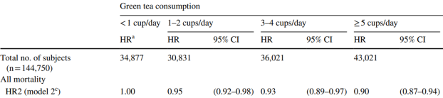
In terms of mortality risk, lowest risk of death for all causes was associated with uric acid levels of 5 – 7 mg/dL for men and 4 – 6 mg/dL for women in the 9,118 adults (average age, 43y) of Hu et al. (2019). Also note the U-shaped curve for both genders, whereas mortality risk increases at both low and high levels of uric acid:

Screen Shot 2020-01-05 at 2.55.00 PM.png

Similarly, the lowest risk of death for all causes was associated with uric acid levels of 7 mg/dL for men, and 4 mg/dL for women in the 375,163 adults (average age, 40y) of Cho et al. (2018), with mortality risk significantly increasing at uric levels < 3.5 and > 9.5 mg/dL for men, < 2.5 and > 7.5 mg/dL for women. Collectively, these 2 studies in middle-aged adults suggest that uric acid levels ~ 4 mg/dL for women and ~7 for men may be optimal for reducing risk of disease for all causes. It’s also important to note that both low and higher values are associated with an increased mortality risk.

The data for the Hu and Chu studies are in younger adults, so how does the data look in older adults? Lowest all-cause mortality risk was associated with uric acid levels between 4 – 5 mg/dL in the 121, 771 older adults (average age, 73y) of Tseng et al. (2018), with mortality risk significantly increasing below 4 and > 8:

Screen Shot 2020-01-05 at 2.43.46 PM.png

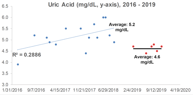
What are my uric acid levels? From 2016 to 2018, I measured it 15x, and although my average value of 5.2 mg/dL is not too low or too high in terms of an increased all-cause mortality risk, it increased during that 3-year period (R2 = 0.2886). When considering that uric acid increases during aging, can I reduce it with diet?

ua ml

Because I track my daily nutritional intake, I can look for correlations between my dietary intake with circulating biomarkers. Interestingly, a moderately strong correlation between my lycopene intake with uric acid (R2 = 0.3343, p=0.024) was present from 2016 to 2018:

ua vs lyco.png

Lycopene is found almost exclusively in tomatoes and watermelon. If these foods are related to my increasing levels of uric acid, if I ate less of them, I’d expect to see a corresponding decrease in uric acid. So, in 2019, I ate less of these foods, thereby reducing my average lycopene intake from 11,585 to 9,132 micrograms per day. How did that affect circulating levels of uric acid?

In 6 measurements for 2019, my average uric acid level was 4.6 mg/dL, a value that was significantly different (p=0.02) from the 2016-2018 average of 5.2 mg/dL. Whether eating less watermelon and tomatoes caused the decrease is unknown, but it’s good to know that uric acid can be potentially modified with dietary change!

my ua


If you’re interested, please have a look at my book!

References

Cho SK, Chang Y, Kim I, Ryu S. U-Shaped Association Between Serum Uric Acid Level and Risk of Mortality: A Cohort Study. Arthritis Rheumatol. 2018 Jul;70(7):1122-1132. doi: 10.1002/art.40472.

Hu L, Hu G, Xu BP, Zhu L, Zhou W, Wang T, Bao H, Cheng X. U-Shaped Association of Serum Uric Acid with All-cause and Cause-Specific Mortality in US Adults: A Cohort Study. J Clin Endocrinol Metab. 2019 Oct 25. pii: dgz068. doi: 10.1210/clinem/dgz068.

Kuzuya M, Ando F, Iguchi A, Shimokata H. Effect of aging on serum uric acid levelslongitudinal changes in a large Japanese population group. J Gerontol A Biol Sci Med Sci. 2002 Oct;57(10):M660-4.

Tseng WC, Chen YT, Ou SM, Shih CJ, Tarng DC; Taiwan Geriatric Kidney Disease (TGKD) Research Group. U-Shaped Association Between Serum Uric Acid Levels With Cardiovascular and All-Cause Mortality in the Elderly: The Role of Malnourishment. J Am Heart Assoc. 2018 Feb 10;7(4). pii: e007523. doi: 10.1161/JAHA.117.007523.

Resting Heart Rate, Heart Rate Variability: December 2019 Update

In earlier posts, I reported year-over-year improvements for my resting heart rate (RHR), from 51.5 (bpm) when I first started tracking in August 2018 to 48 bpm in November 2019 (https://michaellustgarten.wordpress.com/2019/12/05/resting-heart-rate-heart-rate-variability-still-making-progress/). Did my year-over-year RHR improvement continue in December 2019?

As shown below, in December 2018, my average RHR was 49.5 bpm. In December 2019, it was 47.5! These data are significantly different (p=6.5E-05):

rhr dec 2019

While RHR is one metric of cardiovascular health, heart rate variability (HRV) is another. With a stronger heart, the expectation would be a lower RHR, but a higher HRV. December 2019 was my best month ever for HRV, with an average HRV value of 86.3!

hrv 12 2019

Also note that December 2019’s HRV value is significantly different when compared with December 2018 (p=1.6E-11).

How am I able to continuously improve my RHR, and recently, my HRV? I average 15-20 miles of walking per week, and 3-4 days/week of structured exercise (1 hr/session), including a combination of weights, core, and stretching. My average HR during my structured workouts had been ~105 bpm prior to the past few months, but in November and December 2019 I made more of an effort to minimize rest periods, and included higher reps to keep my exercise HR as high as possible. My goal is to get my RHR to 40 bpm, which is associated with maximally reduced risk of death for all causes (https://michaellustgarten.wordpress.com/2019/02/02/resting-heart-rate-whats-optimal/). Stay tuned for more RHR and HRV data next month!

If you’re interested, please have a look at my book!

Epigenetic Aging: Inflammation, Exercise, Smoking

Besides diet (https://michaellustgarten.wordpress.com/2019/12/07/slowing-epigenetic-aging-with-diet/), are there other factors that may impact epigenetic aging? First, let’s have a look at clinically relevant variables, including inflammation, the lipid profile, kidney function, blood pressure, and body size/dimensions (Liu et al. 2019):

EA crp.png

One of the strongest correlations for the clinical variables with epigenetic aging (AgeAccelGrim) is found for C-reactive protein (CRP), with higher CRP being associated with an older epigenetic age. This data supports the hypothesis that CRP levels as low as possible may be representative of biological youth, which I’ve previously written about (https://michaellustgarten.wordpress.com/2019/10/19/optimizing-biological-age-crp/). Similarly, higher values for insulin, glucose, triglycerides, systolic blood pressure, BMI, and the waist/hip ratio were correlated with an older epigenetic age, whereas higher HDL was correlated with a younger epigenetic age. Significant correlations were not identified for total or LDL cholesterol, creatinine, or diastolic blood pressure.

Investigating further, the strongest correlation for epigenetic aging was found for smoking, as current smokers had an older epigenetic age. In contrast, those who exercised, drank alcohol, and that had higher levels of education and income had younger epigenetic ages (Liu et al. 2019):

exerc ea


References

Lu AT, Quach A, Wilson JG, Reiner AP, Aviv A, Raj K, Hou L, Baccarelli AA, Li Y, Stewart JD, Whitsel EA, Assimes TL, Ferrucci L, Horvath S. DNA methylation GrimAge strongly predicts lifespan and healthspan. Aging (Albany NY). 2019 Jan 21;11(2):303-327. doi: 10.18632/aging.101684.

If you’re interested, please have a look at my book!

Epigenetic Aging: Can It Be Slowed With Diet?

Having a faster rate of epigenetic aging, as measured by the epigenetic age metric, AgeAccelGrim, is associated with a significantly increased risk of death for all causes in a variety of cohorts, including the Framingham Heart Study (FHS), the Women’s Health Initiative (WHI) study, the InChianti study, the Jackson Heart Study (JHS), and collectively, when evaluated as a meta-analysis (Lu et al. 2019):

Screen Shot 2019-12-07 at 2.23.27 PM.png

With the goal of minimizing disease risk and maximizing longevity, can epigenetic aging be slowed? Shown below is the correlation between dietary components with AgeAccelGrim. Dietary factors that were significantly associated  (the column labelled, “p”) with a younger epigenetic age were carbohydrate intake, dairy, whole grains, fruit, and vegetables. In contrast, dietary fat intake and red meat were associated with older epigenetic ages (Lu et al. 2019):

Screen Shot 2019-12-07 at 2.34.50 PM.png

Note that dietary recall data as a means for identifying nutrient intake can be unreliable-a better measure of dietary intake is circulating biomarkers. Are there associations between circulating biomarkers of nutrient intake with epigenetic aging?

Higher blood levels of carotenoids, including lycopene, alpha- and beta-carotene, lutein+zeaxanthin, and beta-cryptoxanthin were associated with a younger epigenetic age (Lu et al. 2019):

epi veg

If your goal is optimal health and longevity, eating foods that are rich in these nutrients may be an important strategy for slowing epigenetic aging. Which foods contain these nutrients? Carotenoids are found almost exclusively in vegetables and fruits. For example, lycopene is enriched in watermelon and tomatoes, alpha- and beta-carotene is high in carrots, orange vegetables (sweet potato, squash, pumpkin) and greens, lutein+zeaxanthin is prevalent in greens, and beta-cryptoxanthin’s highest levels are found in butternut squash and red bell peppers.

If you’re interested, please have a look at my book!

References

Nutrient composition data: https://reedir.arsnet.usda.gov/codesearchwebapp/(S(ujsr52ygvp0tw13m1luk0rny))/CodeSearch.aspx

Lu AT, Quach A, Wilson JG, Reiner AP, Aviv A, Raj K, Hou L, Baccarelli AA, Li Y, Stewart JD, Whitsel EA, Assimes TL, Ferrucci L, Horvath S. DNA methylation GrimAge strongly predicts lifespan and healthspan. Aging (Albany NY). 2019 Jan 21;11(2):303-327. doi: 10.18632/aging.101684.

Resting Heart Rate, Heart Rate Variability: Still Making Progress?

Although many of my posts aimed at improving health and longevity are focused on diet, in this post I’ll show data that demonstrates that I’ve been able to steadily improve my cardiovascualr fitness. In earlier posts I reported that a resting heart rate (RHR) of 40 beats per minute (bpm) was associated with lowest risk for all-cause mortality (https://michaellustgarten.wordpress.com/2019/02/02/resting-heart-rate-whats-optimal/), and I noted my own RHR progress in a year-over-year update, from values of 51.5 – 52.7 bpm in August – Sept 2018 to 49.3 – 48.7 bpm during the same months in 2019 (see https://michaellustgarten.wordpress.com/2019/10/08/resting-heart-rate-year-over-year-update/). Have I continued to make progress?

Shown below are 2 more months of RHR data, from August – November 2018, and the data for those months in 2019:
rhr 4 mo
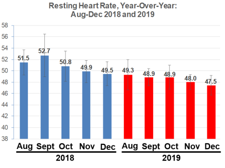
From August – November 2018, I reduced my RHR from ~52 to 50 bpm, whereas in 2019, I made smaller progress, but the trend is still downward, from 49 to 48 bpm. The 2018 data is significantly different from the 2019 data, as assessed by single-factor ANOVA (p = 8E-14).

Adding strength to these findings is that my heart rate variability (HRV), as a second index of cardiovascular health, has increased during the same period:

hrv2
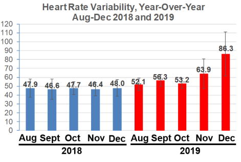
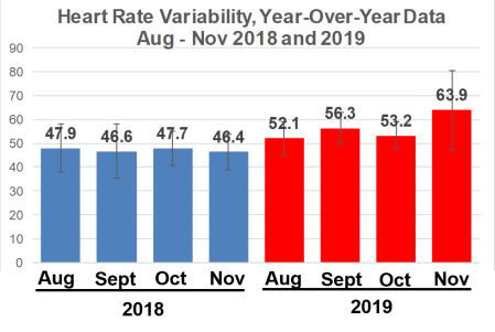
Note that from August – November 2018, my average daily HRV value never topped 48, whereas during the same 4 months in 2019, it was never lower than 52.1, with my best ever HRV values found in November. The 2018 is significantly different when compared with 2019, again based on single-factor ANOVA (p = 5.2E-13).

How am I improving my cardiovascular fitness? That’s a topic for another post, but note that my strength is still pretty good, as evidenced by my 12 pull-ups in the video below!

https://www.youtube.com/watch?v=dLktQvFz70Q

If you’re interested, have a look at my book!

Which Blood Test Analyte Is Most Important For Predicting Biologic Age?

Three studies have investigated the ability of blood test analytes to predict biological age. First, when considering the top 20 variables that were associated with biological age in aging.ai, albumin contributed most to this prediction, almost 2x more than circulating levels of glucose (Mamoshina et al. 2018):

Screen Shot 2019-12-01 at 1.04.59 PM.png

Second, albumin was one of the 9 blood test variables that were best able to predict biological age when using the Phenotypic Age calculator.  However, as shown below, it didn’t come in first place, but fifth. Interestingly, the analyte that contributed most to biological age prediction was the red cell distribution width (RDW%), with glucose again in second place (Levine et al. 2018):

Screen Shot 2019-12-01 at 1.21.58 PM

Third, Earls et al. (2019) used the Klemera-Doubal algorithm (Klemera and Doubal, 2006) in conjunction with blood test data to predict biological age. Regardless if the blood was analyzed by Labcorp or Quest, higher levels of albumin (the left side of both images below) were associated with the greatest reduction in biological age, up to 5 years! In contrast, HbA1c was associated with a higher biological age when measured by Labcorp (top image, right side), and second to lead in the Quest analysis (bottom image, right side). Interestingly, glucose came in third and fifth in the Labcorb and Quest data sets, respectively, in terms of its positive association with biological age.

Screen Shot 2019-12-01 at 12.59.22 PM

Glucose would’ve been an obvious choice, but would you have guessed that albumin may be just as important, and potentially more important for predicting biological age?


If you’re interested, please have a look at my book!

References

Earls JC, Rappaport N, Heath L, Wilmanski T, Magis AT, Schork NJ, Omenn GS, Lovejoy J, Hood L, Price ND. Multi-Omic Biological Age Estimation and Its Correlation With Wellness and Disease Phenotypes: A Longitudinal Study of 3,558 Individuals. J Gerontol A Biol Sci Med Sci. 2019 Nov 13;74(Supplement_1):S52-S60. doi: 10.1093/gerona/glz220.

Klemera P, Doubal S. A new approach to the concept and computation of biological age. Mech Ageing Dev. 2006;127:240–248. doi:10.1016/j. mad.2005.10.004

Levine ME, Lu AT, Quach A, Chen BH, Assimes TL, Bandinelli S, Hou L, Baccarelli AA, Stewart JD, Li Y, Whitsel EA, Wilson JG, Reiner AP, Aviv A, Lohman K, Liu Y, Ferrucci L, Horvath S. An epigenetic biomarker of aging for lifespan and healthspanAging (Albany NY). 2018 Apr 18;10(4):573-591. doi: 10.18632/aging.101414.

Mamoshina P, Kochetov K, Putin E, Cortese F, Aliper A, Lee WS, Ahn SM, Uhn L, Skjodt N, Kovalchuk O, Scheibye-Knudsen M, Zhavoronkov A. Population specific biomarkers of human aging: a big data study using South Korean, Canadian and Eastern European patient populations. J Gerontol A Biol Sci Med Sci. 2018 Jan 11.